

Sophisticated Analytical Instruments Facility
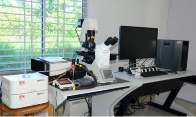
| Name of the equipment/facility: Confocal Laser Scanning Microscopy(CLSM) |  |
||||||||||||||||||||
| Make: Leica Microsystems, Germany | |||||||||||||||||||||
| Model: Leica TCS SP8 | |||||||||||||||||||||
| Specification: | |||||||||||||||||||||
- Bright field, Fluorescence and differential interface contrast (DIC) illumination with facility for confocal scan head attachment
- Automatic (motorized) beam path selection for visual and confocal imaging
- Detectors are capable of working in Intensity and Spectral mode Imaging. Should be capable of simultaneous detection and separation of minimum 5 fluorophores. The detection unit is a combination of 2 PMT & 3 GaAsP/HyD.
- The scan head should be able to perform fast dynamic live cell time lapse imaging with ROI capability with a high speed of 100-300 fps or better @512X32 resolution OR scan speed should be 7-10fps or better @512X512 resolution
- Lasers: Pre aligned Solid State/ Gas State laser module with laser lines of
a) Multi-line Ar laser with 458/488/514nm.
b) DPSS 561 nm
c) HeNe 633 nm
d) Blue Diode Laser 405/408nm.
Microsocpe is equipped with either peizo stage or bidirectional galvo stage for fastest possible Z sections, with travel range of at least 100 microns or better for fast imaging in Confocal and SR mode.
|
|||||||||||||||||||||
| User Instructions: | |||||||||||||||||||||
- User has to bring their samples personally or send the mounted samples on permanent slides suitable to fit in inverted microscope of CLSM
- User has to provide complete sample details with type of tagging or stain, suitable wavelength, need of co-localization, live imaging etc. depending on the application and accordingly the samples will be charged
- Users are encouraged to be present during the analysis of their samples. Users who are interested in specialised experiments are advised to contact Central Instrumentation facility, Bhatnagar Building of CSIR-NEIST, Jorhat-785 006 for an appointment. All users are required to acknowledge the use of the facility when the results are published or presented in a symposium / conference. It will be highly appreciated if users send a Xerox copy of such publication/report to the Central Instrumentation facility, Bhatnagar Building of CSIR-NEIST, Jorhat-785 006.
- User should bring their own DVD/ CD for data transfer. No USB or pen drive allowed.
- All samples should accompany material safety data sheets (MSDS).
|
|||||||||||||||||||||
| Applications: | |||||||||||||||||||||
| Confocal Laser Scanning Microscope is one of the most vital high end equipment to perform wide range of cell biological applications such as co-localization analysis of fluorescently labelled proteins, live 3D imaging, FRET analysis, sub-cellular localization and expression of proteins of plants, bacteria and animal models, in vivo protein-protein interaction assays, detection and analysis of immunolabelled proteins, live video recording of movement of cellular organelles and movement of fluorescently tagged proteins in the cell etc.
|
|||||||||||||||||||||
| Contact | |||||||||||||||||||||
| 1. Dr.Channa Chikkaputtaiah, Senior Scientist,channakeshav@neist.res.in, 8197231132 2. Dr. Jitendra Singh Verma, Scientist, jsverma@neist.res.in, 6000305400 |
|||||||||||||||||||||
| Charges (Excluding Taxes) | |||||||||||||||||||||
Charges (Excluding Taxes):
|
|||||||||||||||||||||